Publication Information
ISSN: 2641-6859
Frequency: Continuous
Format: PDF and HTML
Versions: Online (Open Access)
Year first Published: 2018
Language: English
| Journal Menu |
| Editorial Board |
| Reviewer Board |
| Articles |
| Open Access |
| Special Issue Proposals |
| Guidelines for Authors |
| Guidelines for Editors |
| Guidelines for Reviewers |
| Membership |
| Fee and Guidelines |
 |
Partial Posterior Interosseous Nerve Palsy: A Case Report
Seide Karasel*
Department of Physical Medicine and Rehabilitation, Famagusta State Hospital, Cyprus
Received Date: April 27, 2020; Accepted Date: May 13, 2020; Published Date: May 21, 2020
*Corresponding author: Seide Karasel, Department of Physical Medicine and Rehabilitation, Famagusta State Hospital, Cyprus. Tel: 3923650255; E-mail: karaselseide@yahoo.com
Citation: Karasel S (2020) Partial Posterior Interosseous Nerve Palsy: A Case Report. Adv Ortho and Sprts Med: AOASM-121.
Summary
Compression of the radial nerve at the level of the elbow and forearm; it causes two different clinical pictures as radial tunnel syndrome and posterior interosseous nerve syndrome. Radial tunnel syndrome causes pain and rarely sensory impairment, while motor weakness does not. In posterior interosseous nerve syndrome, hand and finger extensor muscles develop weakness but no sensory impairment is observed. In this article, a patient who developed spontaneous right hand third and fourth finger loss of extension without trauma, without posterior lesion detection was presented, and the importance of considering the partial posterior interosseous nerve syndrome in clinical practice in the differential diagnosis of radial nerve compression and radial division of brachial plexus and the muscles innervated are summarized.
Keywords: Posterior interosseous nerve syndrome; Radial nerve
Introductıon
The radial nerve, which continues from the posterior cord of the brachial plexus, originates from the C5-T1 spinal nerves. The radial nerve flows through the axilla, goes obliquely from the medial of the humerus to the back and goes down in the spiral groove. It innervates the triceps muscle and proceeds through the two heads of the triceps muscle. Then the anconeus muscle; distal to the arm innervates brachioradialis, extensor carpi medialislongus and brevis muscles. It provides forearm and wrist extension, forearm flexion in the neutral position, forearm supination, and thumb extension and abduction. The sensory component of the radial nerve carries cutaneous afferent branches from the arm posterior, forearm and hand [1-4]. The radial nerve is divided into two branches at the level of the forearm, forming the PIN(posterior interosseous nerve) (pure motor) and superficial radial nerve (pure sense). PIN innervates the supinator muscle and passes under this muscle, innervating the extensor digitorumkomminis (EDC), extensor carpi ulnaris (ECU) and extensor digitiminimi (EDM) muscles [2].
The extensor carpi radialis of the radial nerve in the clinic is ,after innervating the longus and brevis muscles, there may be weakness in the extensor muscles of the hand and sometimes pain in the forearm and dorsal side of the hand, the sensation is preserved. PIN compression can be complete or partial. Finger extensors of varying degrees are affected by wrist extensors in partial compression. In this article, a patient with spontaneously developing PIN compression is desciribed and the importance of differential diagnosis is emphasized.
Case Report
A seventy-eight-year-old female patient applied to our clinic with the complaint of not being able to lift the third and fourth fingers of her right hand upwards. The patient, whose complaints started a week ago, does not define any trauma and states that there is no known rheumatic disease. There was no feature in the family history of the patient who had a history of hypertension and gastritis. In the neurological examination of the patient, active extension loss was detected in the third and fourth finger metacarpophalangeal (MP) joint. Wrist, thumb, second and fifth finger extensions were preserved (Figure 1, 2). Superficial and deep sensory examination of the arm, forearm and hand was normal, deep tendon reflexes were normoactive in the upper extremity. In the electroneuromyography (ENMG), the right radial nerve combined muscle action potential amplitude was performed by recording from the right extensor indicis proprius muscle and the motor transmission rate was slowed down. Right median and ulnar nerve transmissions were within normal limits, sensory conduction was normaland also electrophysiological findings were normal in the extensor carpi radialis, brachioradialis, triceps and abductor pollicis longus muscles.
We have planned physiotherapy program with electrical stimulation to extensor of fingers and exercises( range of motion exercices, streching exercises and progressive resistive exercises) .And also a hand-wrist splint for use.After the physiotherapy program, she was better before, can able to do some of daily activities easier.

Figure 1: Finger extension lateral view.
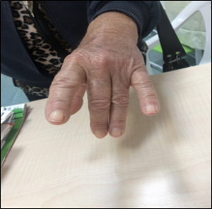
Figure 2: Finger extension, anterior view.
Discussion
The posterior interosseousnerve(PIN) innervates forearm extensor group muscles with 6 branches, which is the motor branch of the radial nerve. The recurrence branch consists of 1, 2, 3 and 4 branches, and the descending branch is 5 and 6 branches. First and second branches of extensor digitorumkomminis (EDC), third branch of extensor carpi ulnaris (ECU), fourth branch of extensor digitiminimi (EDM), fifth branch of extensor index proprius (EIP) and extensor pollicislongus (EPL); the sixth branch innervates the extensor pollicisbrevis (EPB), abductor pollisislongus (APL) and supinator muscles.
There are three types of PIS palsy
Type 1: As a result of recurrent and descending paralysis, all finger extensions are lost.
Type 2: As a result of recurrent paralysis, extension loss occurs in the 3rd, 4th and 5th fingers.
Type 3: Only the descending branch paralysis results in loss of extension in the thumb. EDC, ECU and EDM paralysis, that is type 2 , occurs as a result of a pathology affecting the first, 2nd, 3rd and 4th branches [7]. In our case, the patient with recurrent branch or type 2 PIN paralysis was presented in this study in order to consider it in the clinical differential diagnosis because of its rarity in the literature and to review the PIN again and summarize the muscles it innervates.
In PIN paralysis etiology, systemic diseases such as trauma, brachial neuritis, space-occupying lesion, repetitive overuse of the hand, diabetes mellitus and rheumatoid arthritis, and more rarely, motor neuron disease, multifocal motor neuropathy, hereditary brachial plexopathy, monomelic amyotrophy, Monteggia fracture (proximal fracture of radıus and radial head dislocation) [2]. Type 2: It was emphasized that the loss of extension in the 3rd, 4th and 5th fingers as a result of recurrent branch paralysis and an anatomical variation causing the nerve compression in the distal musculotendinous membrane of the supinatory muscle [7]. Electrodiagnostic studies are important for confirming the diagnosis by localizing nerve damage. It also provides information about the severity of muscle denervation. In addition, X ray and the MRI of the forearm helps to identify the lipoma or other space-occupying lesions that cause trapping by compressing PIS [6].
In our patient, the etiology could not be determined in the examinations performed in terms of Xray, MRI and rheumatological disease. However, in the case, the table was thought to have nerve impingement syndrome due to muscle edema developed as a result of strain of the forearm extensor muscles.
While there was loss of extension in the third and fourth finger of the right hand, the diagnosis of PIN compression was considered primarily because of the extension of the forearm extension and supination, wrist extension, and no loss of sensation, and the diagnosis was confirmed by ENMG. A splint was planned for our patient, medical treatment for muscle edema was arranged and it was decided to be included in the rehabilitation program.
In cases where there is no space-occupying lesion that creates pressure on the nerve in imaging studies in patients with PIN compression; conservative treatment methods such as regulation of activity, splinting, physical therapy, anti-inflammatory drugs and / or corticosteroid injections are recommended. [2, 4] If there is no improvement in the findings within six months, the probability of recovery is lower, and in this case, patients are referred to surgery [2, 4].
Referances
- Mumenthaler M, Stöhr M, Müller-Vahl H, çeviri:Börü Ü (2005) Periferik Sinir Lezyonları ve Radiküler Sendromlar, Nobel Tıp Kitabevleri, 2005: 224-319.
- Bevelaqua AC, Hayter CL, Feinberg JH, Rodeo SA (2012) Posterior Interosseous Neuropathy: Electrodiagnostic Evaluation. HSS J 8: 184-189.
- Ertekin C (2006) Pleksus Brakiyalisten Çıkan Sinirler: Sentral ve Periferik EMG Anatomi-Fizyoloji-Klinik. Meta basım matbaacılık, Bornova-İzmir 2006: 387-453.
- Dumitru D (2002) Focal Peripheral Neuropathies. In: Dumitru D, ed. Electrodiagnostic Medicine. Hanley&Belfus, Philadelphia 2002:1043-126.
- Han BR, Cho YJ, Yang JS, Kang SH, Choi HJ (2014) Clinical features of wrist drop caused by compressive radial neuropathy and its anatomical considerations. J Korean Neurosurg Soc 55: 148-151
- Knutsen EJ, Calfee RP (2013) Uncommon upper extremity compression neuropathies. Hand Clin 29: 443-453.
- Suematsu N (1998) Hirayama T. Posterior interosseous nerve palsy. J Hand Surg Br 23: 104-106.
