Publication Information
Frequency: Continuous
Format: PDF and HTML
Versions: Online (Open Access)
Language: English
| Journal Menu |
| Editorial Board |
| Reviewer Board |
| Articles |
| Open Access |
| Special Issue Proposals |
| Guidelines for Authors |
| Guidelines for Editors |
| Guidelines for Reviewers |
| Membership |
| Fee and Guidelines |
 |
Giant Tonsilloliths Suspected of Tonsillar Cancer
Sørensen RB1*, Amstrup T2, Hvilsom GB1
1Department of Ear, Nose, Throat and Maxillofacial Surgery at Zealand University Hospital, Koege, Denmark
2The Ear-Nose and Throat Clinic, Nykoebing Falster, Denmark
Received Date: January 24, 2023; Accepted Date: January 30, 2023; Published Date: February 04, 2023;
*Corresponding author: Rasmus Blaabjerg Ahm Sørensen. Address: Department of Ear, Nose, Throat and Maxillofacial Surgery at Zealand University Hospital, Lykkebækvej 1, 4600, Koege, Denmark. Telephone: (0045)29896845. E-mail: rasmus.blahm@gmail.com
Citation: Sørensen RB, Amstrup T, Hvilsom GB (2023) Giant Tonsilloliths Suspected of Tonsillar Cancer. Annals of Otorhinolaryngology – Head and Neck: AOHNS-102.
DOI: 10.37722/AOHNS.2023101
Abstract
Introduction: Giant tonsilloliths are rare and only few cases have been described in the literature.
We present a casereport of a 71-year old female with giant tonsilloliths misinterpreted as tonsillar cancer.
Methods: This case was recorded in the Department of Ear, Nose, Throat and Maxillofacial Surgery at Zealand University Hospital, Koege, Denmark. A 71-year old female with giant tonsilloliths misinterpreted as tonsillar cancer. The patient presented with three weeks of odynophagia, most profound in the right side of her mouth and a white area in the lower part of the right tonsil interpreted as a necrotic area. After surgical intervention three tonsilloliths were removed. The patient was contacted 3 months after surgery and reported that she had had no symptoms post-surgery.
Conclusion: Giant tonsilloliths are rare and can be misinterpreted as tonsillar cancer due to the presentation of unilateral symptoms mimicking malignancy.
Keywords: Giant Tonsilloliths; Tonsil Stones; Tonsilloliths; Tonsillar Cancer
Introduction
Tonsilloliths are calcified material accumulated in the tonsils. Tonsilloliths has been known for centuries and is believed first to be described by Lang in 1560 (1) however the pathogenesis remains unknown. It is believed that the concrements are formed by mineralization of accumulates of cellular debris and microorganisms within the tonsillar crypts due to recurrent inflammation of the tonsils (1-4). Small tonsilloliths are common and it is an underdiagnosed condition (5). In a retrospective study of 482 patients Oda et al (5) evaluated CT scans and panoramic radiographs, they observed 46,1 % with tonsilloliths without symptoms from the oropharyngeal area. Giant tonsilloliths are however rare and only few cases have been described in the literature.
We present a case of giant tonsilloliths.
Methods: This case was recorded in the Department of Ear, Nose, Throat and Maxillofacial Surgery at Zealand University Hospital, Koege, Denmark.
Patient information
A 71-year-old female initially referred to the ENT department due to suspicion of tonsillar cancer, presented with a history of three weeks of odynophagia, most profound in the right side of her mouth and a white area in the lower part of the right tonsil interpreted as a necrotic area. The patient did not experience otalgia nor dysphagia. The patient reported recurrent periods of odynophagia through her life and had a history of alcohol overuse and 40 years of tobacco use. The patient’s medical history included non-progressing lung cancer, osteoporosis, intermittent claudication and depression. She did not have a history of gallstone, kidney stone, Wharton’s duct calculi nor recurrent acute tonsilitis.
Clinical findings
Inspecting the oral cavity, a 5 x 10 mm white area was identified in the anterior, lower part of the right tonsil (figure 1). The area of the tonsil was solid on palpation. No pathological finding was observed in the left tonsil. The remaining ENT examination was unremarkable. No imaging was performed.
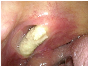
Figure 1: The patient’s right tonsil prior to surgery. A tonsillolith is seen protruding into the oral cavity.
Therapeutic intervention
After administration of infiltration local anaesthetic the solid element was grasped with a pean, and 3 large tonsilloliths were evacuated. Respectively measuring 13 mm x 13 mm, 20 mm x 6 mm and 11 mm x 10 mm (figure 2). To remove the last of the three tonsilloliths a small incision was necessary in the superior part of the right tonsil.
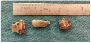
Figure 2: The three tonsilloliths.
Follow-up and outcomes
The patient was instructed to wash her mouth with chlorhexidine mouthwash regularly and contact the department if she had any problems. The patient was contacted 3 months later and reported that she had had no symptoms.
Discussion
We report a case of a 71-year-old female referred to our ENT department due to suspicion of a right sided tonsillar cancer, fortunately 3 tonsilloliths were removed without suspicion of malignancy. Giant tonsilloliths can mimic malignancy by presenting with sudden unilateral odynophagia and a white firm area in the palatine tonsil, which can be misinterpreted as a necrotic region both on observation and palpation. Furthermore, the patient had a history of lung cancer, a long history of tobacco use and alcohol overuse disposing to malignancy. Few other cases of giant tonsilloliths misinterpreted as malignancy has been reported (4,6,7).
Tonsilloliths are the most common type of calcification of the soft tissue in the head and neck region. The condition is underdiagnosed since it is often either asymptomatic or present with vague, non-specific symptoms such as halitosis (8)(3). Other symptoms associated with tonsilloliths are sore throat (1,3,4,9-11), swelling in the tonsillar fossa (1,3,4,9,12), dysphagia (4,11–13), odynophagia (1,4,6,7,10,13), otalgia (1,4,7,9,13), peritonsillar abscess (14), swelling in the submaxillary triangle (4,15), foreign body sensations (6,12), irritable cough (15) and recurrent throat infection (3,11,12,14,15). All unspecific symptoms seen in many of our patients on a daily basis in an ENT department.
The age of patient reported with tonsilloliths range between under 9 years to over 90 years (3-5,16) though there is an increase in prevalence with age, especially over 40 years of age (5,8,17,18). It is often an incidental finding on routine radiographic examinations (5). Most studies suggest that tonsilloliths are more frequent in the male population and more often unilateral rather than bilateral (8,16,17) however, there is no consensus in the literature (4,5). Patients often have a history of tonsillectomy, gallstones, kidney stones or Wharton’s duct calculi (15). This was not the case in our patient.
Examining the tonsilloliths most of the stones consist of calcium carbonate and calcium phosphate, but other minerals have also been reported (1). They are usually no more than 3-4 mm in length (5) and can be different in shape and colour (15).Differential diagnosis of tonsilloliths include foreign body, calcified granulomas, tonsillar malignancy, enlarged styloid processes, and embryonic rests originating from the branchial arches (3).
Tonsilloliths can be identified on CT scans. Cone Beam CT has proven to be a very sensitive tool in diagnosing tonsilloliths, whereas panoramic radiographs are less useful (5)(8). Cho et al (9) report ultrasound as a diagnostic tool which is easily accessible in every ENT department in Denmark.
Conclusion
Tonsilloliths are not uncommon, and are often asymptomatic or present with very vague symptoms. Giant tonsilloliths though are rare and can be misinterpreted as tonsillar cancer due to the presentation of unilateral symptoms mimicking malignancy.
Acknowledgments: This study would like to thank the Department of Ear, Nose, Throat and Maxillofacial Surgery at Zealand University Hospital, Koege, Denmark.
Declaration of conflicting interests: The Authors declare that there is no conflict of interest.
Funding: This research received no specific grant from any funding agency in the public, commercial, or not-for-profit sectors.
References
- Kanotra S, Kanotra S, Paul J. A giant tonsillolith. Indian J Otolaryngol Head Neck Surg [Internet] 2008 Sep; 60:277-80.
- Gadgil RM. An unusually large tonsillolith. Oral Surgery, Oral Medicine, Oral Pathology 1984 Aug;58.
- Alfayez A, Albesher MB, Alqabasani MA. A giant tonsillolith. Saudi Med J [Internet] 2018 Apr; 39:412-4.
- Mesolella M, Cimmino M, di Martino M, Criscuoli G, Albanese L, Galli V. Tonsillolith. Case report and review of the literature. Acta otorhinolaryngologica Italica : organo ufficiale della Societa italiana di otorinolaringologia e chirurgia cervico-facciale 2004 Oct;24.
- Oda M, Kito S, Tanaka T, Nishida I, Awano S, Fujita Y, et al. Prevalence and imaging characteristics of detectable tonsilloliths on 482 pairs of consecutive CT and panoramic radiographs. BMC Oral Health [Internet] 2013 Oct 14; 13:54.
- Chan J, Rashid M, Karagama Y. An unusual case of a tonsillolith. Case Rep Med 2012;2012.
- Revel MP, Bely N, Laccourreye O, Naudo P, Hartl D, Brasnu D. Giant tonsillolith. Ann Otol Rhinol Laryngol 1998 Mar;107.
- Yalcin ED, Ararat E. Prevalence of soft tissue calcifications in the head and neck region: A cone-beam computed tomography study. Niger J Clin Pract [Internet] 2020 Jun; 23:759-63.
- Cho W, Park H. Transoral sonographic diagnosis of tonsilloliths: report of 3 cases. Journal of ultrasound in medicine 2013; 32:2037-42.
- Shikino K, Ikusaka M. Tonsillolith. Clin Case Rep 2021 Jun;9.
- Thakur JS, Minhas RS, Thakur A, Sharma DR, Mohindroo NK. Giant tonsillolith causing odynophagia in a child: a rare case report. Cases J 2008 Jul 18;1.
- Silvestre-Donat FJ, Pla-Mocholi A, Estelles-Ferriol E, Martinez-Mihi V. Giant tonsillolith: report of a case. Medicina oral, patologia oral y cirugia bucal 10.
- Kim KS. Referred otalgia induced by a large tonsillolith. Korean J Fam Med 2013 May;34(3).
- Kimura H, Ohashi N, Nakagawa H, Asai M, Koizumi F. Large tonsillolith mimicking peritonsillar abscess: a case report Auris Nasus Larynx. 1993;20.
- Siber S, Hat J, Brakus I, Biočić J, Brajdić D, Zajc I, et al. Tonsillolithiasis and orofacial pain. Gerodontology [Internet] 2012 Jun;29: e1157-60.
- Ram S, Siar CH, Ismail SM, Prepageran N. Pseudo bilateral tonsilloliths: a case report and review of the literature. Oral Surg Oral Med Oral Pathol Oral Radiol Endod 2004 Jul;98.
- Khojastepour L, Haghnegahdar A, Sayar H. Prevalence of Soft Tissue Calcifications in CBCT Images of Mandibular Region. Journal of dentistry (Shiraz, Iran) 2017 Jun;18.
- Garay I, Netto HD, Olate S. Soft tissue calcified in mandibular angle area observed by means of panoramic radiography. Int J Clin Exp Med 2014;7.