Publication Information
ISSN: 2688-0873
Frequency: Continuous
Format: PDF and HTML
Versions: Online (Open Access)
Year first Published: 2018
Language: English
| Journal Menu |
| Editorial Board |
| Reviewer Board |
| Articles |
| Archives |
| Open Access |
| Special Issue Proposals |
| Guidelines for Authors |
| Guidelines for Editors |
| Guidelines for Reviewers |
| Membership |
| Fee and Guidelines |
 |
Exoscopes, a new dimension in Neurosurgery?
Rene Regatschnig1, 2*, Eva Himmelbauer1, 2, Stefan Schweiger1, 2, Nicola Busse1, 2, Johannes Burtscher1, 2
1Clinical Department of Neurosurgery, State Hospital, Wiener Neustadt, Austria
2Faculty of Medicine and Dentistry, Danube Private University, Krems, Austria
Received Date: February 06, 2024; Accepted Date: February 16, 2024; Published Date: March 06, 2024;
*Corresponding author: R. Regatschnig, Clinical Department of Neurosurgery, State Hospital, Wiener Neustadt, Austria; Email: Rene.Regatschnig@wienerneustadt.lknoe.at
Citation: Regatschnig R, Himmelbauer E, Schweiger S, N. Busse, Burtscher J; Exoscopes, a new dimension in Neurosurgery? (2024). Jr Surg Opetech Anesthesia: JSOPA-118
DOI: 10.37722/JSOTA.2024102
Abstract
Objective: We tested 4 different exoscopes and compared them to each other and to the operating microscope (OM) to evaluate if exoscopes can replace OM in Neurosurgery at present time.
Methods: The exoscopes were tested in the lab and in the operating room. The systems were available for testing for several weeks at our department. We compared the exoscopes to each other and to the OM. The exoscopes were used in standard cranial and spinal procedures. All Neurosurgeons of the department had the chance to use the exoscopes and to evaluate them.
Results: Each surgeon in the department was trained in the lab to use the exoscopes. After each procedure, the exoscope was evaluated by the surgeon with a questionnaire. Overall, the exoscopes achieved better scores than the OM, especially regarding ergonomics, visualization, and viewing angles. With the training in the lab, there was a shallow learning curve. None of the attending neurosurgeons switched back to the OM during any procedure. Nevertheless, there were significant differences between the different exoscopes.
Conclusion: After testing the exoscopes in different neurosurgical procedures, we decided to replace our OM with an exoscope. In our opinion, the advantages of the exoscopes outweigh the drawbacks. We are convinced that over time and with future developments, exoscopes will replace OM and will add a new dimension in neurosurgical practice.
Keywords: Exoscope; operating microscope; optical quality, ergonomics
Introduction
Operating microscopes (OM) have been the workhorse for neurosurgical imaging since the late 1960s and have led to new neurosurgical procedures, new techniques, and instruments through improved visualization, better illumination, and magnification of the operating field (1–4).
Over the years, OM was improved, and additional features were added, like the integration of neuronavigation and fluorographic imaging for oncologic and vascular neurosurgery (5–11). In recent years, exoscopes were developed and introduced to neurosurgical practice as an alternative to the established OM (12–23). Compared to microscopes, which are analog systems by design, exoscopes are digital systems. Currently available exoscopes share some common features, but also show differences among them. The images created bz exoscopes are presented via monitors to the surgeon and the Operating room staff. Exoscopes have already demonstrated their usefulness in different surgical specialties, such as ENT, general surgery, plastic surgery, and orofacial surgery (23–32).
Material and methods
We tested 4 different exoscopes, for several weeks each, at our department. Before operating with the exoscope, we offered serval days of practice with the particular exoscope in the lab. The 4 tested exoscopes were the RoboticScope® (RS; BHS Technologies GmbH, Innsbruck, Austria), Aesculap Aeos® (Braun, Meslungen, Germany) the VITOM® 3D (Karl Storz SE & Co. KG, Tuttlingen, Germany) and the ORBEYE (Olympus, Tokyo, Japan). The goal was to evaluate if Exoscopes can at this time replace a neurosurgical operating microscope. All tested exoscopes share some common features but differ in other features. Compared to the OM, all exoscopes are fully digital systems with digital picture processing and all of them use an LED light source. Eeoscopes use monitors, instead of oculars, to display the image.
The RoboticScope uses a head-mounted display with 2 screens in front of the surgeon´s eye to generate a 3-dimensional (3D) image. The camera is mounted on a robotic arm, which is controlled by specified movements of the surgeon’s head. The functions of the RoboticScope are controlled with a footswitch and by head movement. It is a true hands-free system. The RoboticScope does not feature intraoperative fluorescence.
The Vitom system is based on an endoscopic system. The camera is mounted on a holding arm, which is moved manually. Functions of Vitom are controlled by jog wheel. Vitom 3D does not offer intraoperative fluorescence.
The camera of the Aeos exoscope is mounted on a robotic arm, which can be controlled by footswitch or via 2 separate keypads mounted on each of the handgrips of the unit. It offers intraoperative fluorescence for oncologic and vascular procedures.
The camera of the Orbeye system is a semi-robotic system, which can be controlled single handedly. It is equipped with intraoperative fluorescence for oncologic and vascular procedures. Optical functions can be controlled using a footswitch.
Vitom, Aeos and Orbeye use a 3D screen and polarized glasses for 3D-viewing. None of the exoscopes can be connected to a neuronavigation system.
After each procedure, the respective surgeon filled out a questionnaire which included picture quality, illumination, ergonomics, additional features when used, eyestrain, magnification and whether the surgeon would use the exoscope for this procedure again.
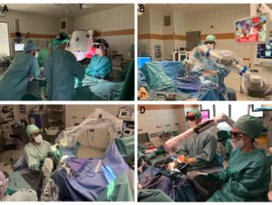
Figure 1: A) RoboticScope, during spinal procedure using head-mounted displays, B) Aeos, displaying possible extreme viewing angles in posterior fossa surgery, C) Vitom 3D, in convexity meningioma surgery, D) Orbeye, in skull base meningioma surgery
Results
Every surgeon of the department had the chance to test every exoscope in the lab and in the operating room. With all exoscopes, we performed standard cranial and spinal neurosurgical procedures. The surgeons were free to use the exoscopes for any procedure. During our test, no surgeon converted back to the operating microscope. The only procedure we did not perform with an exoscope during the period of testing the systems were vascular cases. With the practice in the lab the learning curve was shallow. In the lab, we tried to figure out the best position of the exoscope and the monitors for the different procedures. Nevertheless, in some cases, we had to reposition the exoscope or the monitors intraoperatively. The operating time was equal to procedures performed with the OM. There were no complications related to the use of the exoscope. We did not experience any technical failure of any of the systems. According to the questionnaire, the satisfaction with image quality, magnification and illumination was higher with exoscopes than with microscopes. In some procedures, image quality was an issue. However, most of the time the problem could be resolved by repositioning the monitor. The position of the monitor played a crucial role in picture quality. There was a direct relation between eyestrain and picture quality. Ergonomics was rated high regardless of the operation. There was no surgeon who would not operate the same procedure again using an exoscope. Overall, the surgeons felt more comfortable with the exoscope during cranial than spinal procedures.
The RoboticScope provides good picture quality, and the control of the robotic arm with head movement works well. The main drawback of this system is the weight of the helmet-mounted displays for longer procedures, and the lack of fluorographic imaging. Due to the helmet-mounted displays, situational awareness is reduced, and some surgeons reported that they lost orientation during the procedure. The Vitom 3D is user-friendly and easy to use. It only features digital zoom, which is a drawback. Due to its small size, it works well for superficial pathologies. The Aeos is most comparable to an OM. By controlling the Exoscope with a footswitch there is no need to control the Exoscope by hand. Particularly useful is the implementation of 5-ALA fluorographic imaging, where the surgeon can digitally blend imaging modalities to operate under white light and fluorescence simultaneously. The lock-on-target function improves ergonomics significantly. The picture quality of the orbeye is excellent. The semi-robotic arm made it difficult to position the Orbeye in some situations. Fluoroscopic imaging with Orbeye worked equally well as with OM.
Discussion
This studies confirms that, when compared to the OM, picture quality and depth of field of exoscopes were at least equivalent, which has been confirmed by serval studies (13–15, 18, 33, 34). Digital image processing allows for adapting of the image to the situation and to the surgeon’s preference. The ergonomics of all exoscopes were rated higher compared to the OM, which has been reported in several previous publications (35–41). All tested exoscopes allow for greater viewing angles, which may allow for smaller approaches. LED light sources used in exoscopes are an improvement compared to the Xenon light of OM. Using LED light, we experienced less radiating heat and thus less drying of the tissue within the operating field. The visualization of the operating field on a 3D screen may also improve education (42–44). The missing integration of neuronavigation is a clear drawback of exoscopes compared to OM. Ratings for cranial procedures were higher than for spinal procedures.
Conclusions
After testing several exoscopes, we conclude that in our opinion, the advantages of available exoscopes already outweigh the disadvantages. The digital nature of image processing allows for further development and improvements. We are convinced that Exoscopes will replace OM in the future. With integration of neuronavigation, improvement in user interfacing and further improvement in visual quality, exoscopes may replace more OM in the next couple of years.
Disclosure: No conflict of interest
References
- Kriss TC, Kriss VM. History of the Operating Microscope: From Magnifying Glass to Microneurosurgery. Neurosurgery. 1998 Apr 1; 42(4):899–907.
- Langer DJ, White TG, Schulder M, Boockvar JA, Labib M, Lawton MT. Advances in Intraoperative Optics: A Brief Review of Current Exoscope Platforms. Operative Neurosurgery. 2020 Jul; 19(1):84–93.
- Yaşargil MG, Krayenbühl H. The use of the binocular microscope in neurosurgery. Bibl Ophthalmol. 1970; 81:62–5.
- Uluç K, Kujoth GC, Başkaya MK. Operating microscopes: past, present, and future. Neurosurg Focus. 2009 Sep; 27(3):E4.
- Shurkhay VA, Goryaynov SA, Aleksandrova E V., Spallone A, Potapov AA. Navigation systems in neurosurgery. Voprosy neirokhirurgii imeni NN Burdenko. 2016; 80(6):107.
- Stummer W, Novotny A, Stepp H, Goetz C, Bise K, Reulen HJ. Fluorescence-guided resection of glioblastoma multiforme utilizing 5-ALA-induced porphyrins: a prospective study in 52 consecutive patients. J Neurosurg. 2000 Dec; 93(6):1003–13.
- Hamamcıoğlu MK, Akçakaya MO, Göker B, Kasımcan MÖ, Kırış T. The use of the YELLOW 560nm surgical microscope filter for sodium fluorescein-guided resection of brain tumors: Our preliminary results in a series of 28 patients. Clin Neurol Neurosurg. 2016 Apr; 143:39–45.
- Jhawar SS, Kato Y, Oda J, Oguri D, Sano H, Hirose Y. FLOW 800-assisted surgery for arteriovenous malformation. Journal of Clinical Neuroscience. 2011 Nov; 18(11):1556–7.
- Fukuda K, Kataoka H, Nakajima N, Masuoka J, Satow T, Iihara K. Efficacy of FLOW 800 with Indocyanine Green Videoangiography for the Quantitative Assessment of Flow Dynamics in Cerebral Arteriovenous Malformation Surgery. World Neurosurg. 2015 Feb; 83(2):203–10.
- Dashti R, Laakso A, Niemelä M, Porras M, Hernesniemi J. Microscope-integrated near-infrared indocyanine green videoangiography during surgery of intracranial aneurysms: the Helsinki experience. Surg Neurol. 2009 May; 71(5):543–50.
- Haberland N, Ebmeier K, Hliscs R, Steenbeck J, Grunewald JP, Nowak H, et al. Neuronavigation in surgery of intracranial and spinal tumors. J Cancer Res Clin Oncol. 2000 Aug 10; 126(9):529–41.
- Roberts DW, Strohbehn JW, Hatch JF, Murray W, Kettenberger H. A frameless stereotaxic integration of computerized tomographic imaging and the operating microscope. J Neurosurg. 1986 Oct; 65(4):545–9.
- Montemurro N, Scerrati A, Ricciardi L, Trevisi G. The Exoscope in Neurosurgery: An Overview of the Current Literature of Intraoperative Use in Brain and Spine Surgery. J Clin Med. 2021 Dec 31; 11(1):223.
- Amoo M, Henry J, Javadpour M. Beyond magnification and illumination: preliminary clinical experience with the 4K 3D ORBEYETM exoscope and a literature review. Acta Neurochir (Wien). 2021 Aug 2; 163(8):2107–15.
- Maurer S, Prinz V, Qasem LE, Lucia KE, Rösler J, Picht T, et al. Evaluation of a Novel Three-Dimensional Robotic Digital Microscope (Aeos) in Neurosurgery. Cancers (Basel). 2021 Aug 25; 13(17):4273.
- Ricciardi L, Chaichana KL, Cardia A, Stifano V, Rossini Z, Olivi A, et al. The Exoscope in Neurosurgery: An Innovative “Point of View”. A Systematic Review of the Technical, Surgical, and Educational Aspects. World Neurosurg. 2019 Apr; 124:136–44.
- Hafez A, Elsharkawy A, Schwartz C, Muhammad S, Laakso A, Niemelä M, et al. Comparison of Conventional Microscopic and Exoscopic Experimental Bypass Anastomosis: A Technical Analysis. World Neurosurg. 2020 Mar; 135:e293–9.
- Beez T, Munoz-Bendix C, Beseoglu K, Steiger HJ, Ahmadi SA. First Clinical Applications of a High-Definition Three-Dimensional Exoscope in Pediatric Neurosurgery. Cureus. 2018 Jan 24;
- Rossmann T, Veldeman M, Nurminen V, Huhtakangas J, Niemelä M, Lehecka M. 3D Exoscopes are Noninferior to Operating Microscopes in Aneurysm Surgery: Comparative Single-Surgeon Series of 52 Consecutive Cases. World Neurosurg. 2023 Feb; 170:e200–13.
- Hafez A, Haeren RHL, Dillmann J, Laakso A, Niemelä M, Lehecka M. Comparison of Operating Microscope and Exoscope in a Highly Challenging Experimental Setting. World Neurosurg. 2021 Mar; 147:e468–75.
- Iqbal J, Covell MM, Jabeen S, Nadeem A, Malik Gunjial H, Abdus Saboor H, et al. Comparative analysis of exoscope-assisted spine surgery versus operating microscope: A systematic review. World Neurosurg X. 2024 Jan; 21:100258.
- Murai Y, Sato S, Yui K, Morimoto D, Ozeki T, Yamaguchi M, et al. Preliminary Clinical Microneurosurgical Experience With the 4K3-Dimensional Microvideoscope (ORBEYE) System for Microneurological Surgery: Observation Study. Operative Neurosurgery. 2019 Jun; 16(6):707–16.
- SHIMIZU T, TOYOTA S, NAKAGAWA K, MURAKAMI T, MORI K, KISHIMA H, et al. Retrosigmoid Approach in the Supine Position Using ORBEYE: A Consecutive Series of 14 Cases. Neurol Med Chir (Tokyo). 2020; 61(1):55–61.
- Gandhi S, Tayebi Meybodi A, Belykh E, Cavallo C, Zhao X, Syed MP, et al. Survival Outcomes Among Patients With High-Grade Glioma Treated With 5-Aminolevulinic Acid–Guided Surgery: A Systematic Review and Meta-Analysis. Front Oncol. 2019 Jul 17; 9.
- Hadjipanayis CG, Stummer W. 5-ALA and FDA approval for glioma surgery. J Neurooncol. 2019 Feb 14; 141(3):479–86.
- Festa BM, Zuppardo J, Costantino A, Ferreli F, Spriano G, Mercante G, et al. High-definition 3D exoscope-assisted tonsillectomy. Am J Otolaryngol. 2023 Jan; 44(1):103674.
- Picart T, Berhouma M, Dumot C, Pallud J, Metellus P, Armoiry X, et al. Optimization of high-grade glioma resection using 5-ALA fluorescence-guided surgery: A literature review and practical recommendations from the neuro-oncology club of the French society of neurosurgery. Neurochirurgie. 2019 Aug; 65(4):164–77.
- Ma L, Fei B. Comprehensive review of surgical microscopes: technology development and medical applications. J Biomed Opt. 2021 Jan 4; 26(01).
- Garcia JP, Avila FR, Torres RA, Maita KC, Borna S, Rinker BD, et al. Evaluating the exoscope as an alternative to the operating microscope in plastic surgery. Journal of Plastic, Reconstructive & Aesthetic Surgery. 2023 Oct; 85:376–86.
- Kullar P, Tanna R, Ally M, Vijendren A, Mochloulis G. VITOM 4K 3D Exoscope: A Preliminary Experience in Thyroid Surgery. Cureus. 2021 Jan 14;
- Visocchi M, Mattogno P, Ciappetta P, Barbagallo G, Signorelli F. Combined transoral exoscope and OArm-assisted approach for craniovertebral junction surgery: Light and shadows in single-center experience with improving technologies. J Craniovertebr Junction Spine. 2020; 11(4):293.
- Ferlito S, La Mantia I, Caruso S, Cammaroto G, Chiesa-Estomba CM, Iannella G, et al. High Definition Three-Dimensional Exoscope (VITOM 3D) in E.N.T. Surgery: A Systematic Review of Current Experience. J Clin Med. 2022 Jun 23; 11(13):3639.
- Muhammad S, Lehecka M, Niemelä M. Preliminary experience with a digital robotic exoscope in cranial and spinal surgery: a review of the Synaptive Modus V system. Acta Neurochir (Wien). 2019 Oct 22; 161(10):2175–80.
- Hines K, Hughes LP, Franco D, Sharan AD, Wu C. Exoscope improves visualization and extent of hippocampal resection in temporal lobectomy. Acta Neurochir (Wien). 2022 Nov 8; 165(1):259–63.
- Lavé A, Gondar R, Demetriades AK, Meling TR. Ergonomics and musculoskeletal disorders in neurosurgery: a systematic review. Acta Neurochir (Wien). 2020 Sep 23; 162(9):2213–20.
- Gadjradj PS, Ogenio K, Voigt I, Harhangi BS. Ergonomics and Related Physical Symptoms Among Neurosurgeons. World Neurosurg. 2020 Feb; 134:e432–41.
- Gabrovsky N, Petrov M, Ilkov P, Iordanova I, Velinov N. Subjective workload measurement of the transition from a conventional operative microscope to a Robotic Digital Microscope. A pilot study. Brain and Spine. 2022; 2:100928.
- Raheja A, Mishra S, Garg K, Katiyar V, Sharma R, Tandon V, et al. Impact of different visualization devices on accuracy, efficiency, and dexterity in neurosurgery: a laboratory investigation. Neurosurg Focus. 2021 Jan; 50(1):E18.
- Steinhilber B, Conte L, Seibt R, Herlan S, Tatagiba M, Ebner FH. Musculoskeletal demands in microsurgery—an explorative study comparing the ergonomics of microscope and 3D exoscope. Neurosurg Rev. 2023 Jul 4; 46(1):164.
- Abramovic A, Demetz M, Krigers A, Bauer M, Lener S, Pinggera D, et al. Surgeon’s comfort: The ergonomics of a robotic exoscope using a head-mounted display. Brain and Spine. 2022; 2:100855.
- Demetz M, Abramovic A, Krigers A, Bauer M, Lener S, Pinggera D, et al. Cadaveric study of ergonomics and performance using a robotic exoscope with a head-mounted display in spine surgery. J Robot Surg. 2024 Jan 10; 18(1):6.
- Sack J, Steinberg JA, Rennert RC, Hatefi D, Pannell JS, Levy M, et al. Initial Experience Using a High-Definition 3-Dimensional Exoscope System for Microneurosurgery. Operative Neurosurgery. 2018 Apr; 14(4):395–401.
- Layard Horsfall H, Mao Z, Koh CH, Khan DZ, Muirhead W, Stoyanov D, et al. Comparative Learning Curves of Microscope Versus Exoscope: A Preclinical Randomized Crossover Noninferiority Study. Front Surg. 2022 Jun 6; 9.
- Giammalva GR, Paolini F, Meccio F, Giovannini EA, Provenzano A, Bonosi L, et al. Assessing the Training in Neurosurgery with the Implementation of VITOM-3D Exoscope: Learning Curve on Experimental Model in Neurosurgical Practice. Brain Sci. 2023 Oct 2; 13(10):1409.
