Publication Information
ISSN: 2641-6859
Frequency: Continuous
Format: PDF and HTML
Versions: Online (Open Access)
Year first Published: 2018
Language: English
| Journal Menu |
| Editorial Board |
| Reviewer Board |
| Articles |
| Open Access |
| Special Issue Proposals |
| Guidelines for Authors |
| Guidelines for Editors |
| Guidelines for Reviewers |
| Membership |
| Fee and Guidelines |
 |
Complete Resolution of Extensive Labral tear and Para-labral Cysts on MRI Following 3D Collagen-Based Stem Cell Therapy in Dysplastic Hip: Case Report
Hassan Mubark*
Rheumatologist, Institution: Auckland Regenerative Clinic, Ormiston Specialist Centre, 125 Ormiston Road
Flat Bush, Auckland 2019, New Zealand
Received Date: November 12, 2021; Accepted Date: November 17, 2021; Published Date: November 23, 2021
*Corresponding author: Hassan Mubark, Rheumatologist, Institution: Auckland Regenerative Clinic, Ormiston Specialist Centre, 125 Ormiston Road, Flat Bush, Auckland 2019, New Zealand. Tel: +64 9 2713305/+64 21843513; Fax: +64 9 2770769; Email: drhassanmubark@gmail.com
Citation: Mubark H (2021) Complete Resolution of Extensive Labral tear and Para-labral Cysts on MRI Following 3D Collagen-Based Stem Cell Therapy in Dysplastic Hip: Case Report: Case Report. Adv Ortho and Sprts Med: AOASM-157.
DOI: 10.37722/AOASM.2021701
Abstract
The common causes of adult chronic labral tear(s) include repetitive trauma, usually in elite athletes, capsular hip hypermobility, femoroacetabular impingement, acetabular dysplasia, and in association with degeneration hip osteoarthritis. Labral tears have been well documented in people with hip dysplasia, as this changes the position of the femoral head within the acetabulum. A labral tear can be associated with chondral injury and the risk of developing premature osteoarthritis.
With the negligible penetration of vessels into the labrum, some authors have concluded that no labrum area has the ability for repair, and no clinical studies to date show that labral tears do heal. A surgical choice is the only available option by debriding and repairing labral tears and associated structural defects. We report a thirty-Two-year male lawyer with an athletic background who suffered from right acetabular dysplasia that led to extensive labral tears and para-labral cysts. He was treated with experimental 3D expanded stem cell culture using FDA-approved collagen type1 that crossed-linked with hyaluronic acid, followed by corrective osteotomy to his right hip one month after the stem cell therapy trying to prevent future hip arthroplasty. He underwent post-treatment physical rehabilitation. He reported an outstanding clinical and MRI radiological response in his right hip with the extensive labral tear that has entirely resolved together with the disappearance of the para-labral cysts five and a half months after the 3D stem cell therapy; he was able to return to exercise comfortably with regular exer-cycling and walking. This case stands for a world unique successful clinical and radiological outcome following experimental 3D stem cell culture with the benefit of adding an extra-cellular matrix to the treatment with an enormous potential for non-surgical tissue repair and regeneration of the joint structures. We highly recommend randomized controlled trials to show if those results are consistent.
Keywords: Labral tear; Para-labral cysts; 3D stem cell; Osteoarthritis; healing; Collagen; Acetabular dysplasia
Introduction
The hip labrum has several functions, including joint lubrication, shock absorption, pressure provision, and aiding joint stability [1]. The acetabulum covers around 170° of the femoral head [2]. The acetabular labrum is a fibro-cartilaginous structure that forms the acetabular outlet [3].
Tears of the acetabular labrum commonly occur from two possible processes: a single, acute significant injury, especially involving strenuous hip flexion resistance during kicking or running, or repetitive microtrauma in a hip with chronic, degenerative bony abnormalities [4].
Labral tears often go undiagnosed during an extended period. Evaluation usually begins with a simple X-ray to assess for acetabular dysplasia, osteoarthritis, and other causes of hip pain. While MRI and CT scans are helpful in the diagnosis of labral tear, magnetic resonance arthrography is the diagnostic test of choice, with arthroscopy being the gold standard. Through this case, we prove the complete healing of an extensive labral tear and resolution of para-labral cysts by using experimental collagen-based 3D expanded stem cell therapy injected into both intra-articular and directly into the labral areas.
Case Report
A thirty-two-year-old male, otherwise healthy lawyer. He usually is active in hiking, mountain biking, and skiing. He saw an orthopedic surgeon for an assessment and management.
He developed right hip pain for one year, which initially came with running-based activities. The pain slowly worsened over time, which led to stopping his running. He had significant pain with prolonged walking and prolonged skiing. Examination revealed a range of motion from 0-100° of flexion with 35° of internal rotation and 60° of external rotation in flexion. Pelvis x-ray shows a lateral centre edge angle of 17° on the right with a Tönnis roof angle of 13° on the right. On the left side, the lateral centre edge angle measures 28° with a Tönnis roof angle of 8°. MRI scans show extensive labral tearing of the anterosuperior, lateral and posterosuperior labrum. There is a degree of chondral thinning at the peripheral acetabulum, a small area over the anterior femoral head, and a small para-labral cyst (Figures 1a and 1b). His diagnosis was symptomatic right acetabular dysplasia, extensive labral tearing right hip, and early chondral damage of the right hip.
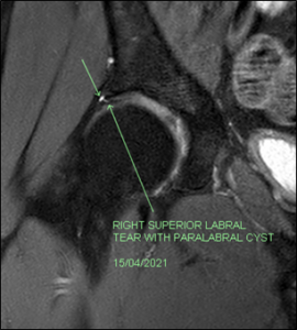
Figure 1a: Pre-treatment coronal MRI of right hip demonstrating extensive labral tear
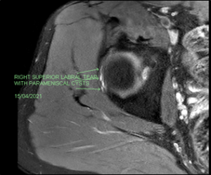
Figure 1b: Pre-treatment axial MRI of right hip demonstrating labral tear multiple labral cysts
He was given the options of conservative treatment versus the surgical choice. During the patient’s research, he wanted to try experimental therapy for both hips, particularly to the right side, as the left hip has mild symptoms compared to the right one. Our regenerative clinic explained a new 3D stem cell therapy trial using collagen1-hyaluronic acid hydrogel to add an extra-cellular matrix for stem cell potentiation and differentiation. After going through the pros and cons with the safety data of our FDA-approved bovine type1 collagen, he was happy to proceed with the therapy. The plastic surgeon performed the abdominal fat harvesting at our hospital. The stem cells were sterilized and cultured for a few weeks to a count of 1 x 10⁸ plus some added cells to culture in 3D hydrogel created by type1 collagen. We cross-linked it with the hyaluronic acid at our specialized laboratory to form good stem cell growth and spheroid masses (Figure 2).
We implanted the stem cells in 3D culture together with platelet-rich plasma in a total fluid of 10 MLS directly into the labrums and hip joints under our office ultrasound guidance using the curved C60 curved probe “Sonosite machine.”
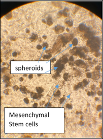
Figure 2: 3D Mesenchymal Stem Cell culture demonstrating Cell growth & Spheroids
The patient decided to go for right hip extra-articular osteotomy to shape the acetabulum as a trial of preserving the hip; this was done successfully one month after the 3D stem cell therapy without interfering with the stem cell therapy.
The patient reported a good clinical outcome and could walk and cycle without deep-seated joint pains. The repeat MRI was done five and a half months post 3D stem cell therapy revealed right hip complete repair of the extensive labral tear and full resolution of the para-labral cysts (Figures 3a, b and c).
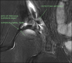
Figure 3a: post-treatment coronal MRI demonstrating healing of the labral tear
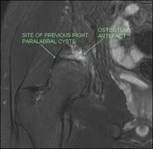
Figure 3b: post-treatment coronal MRI Demonstrating resolution of the para-labral cysts
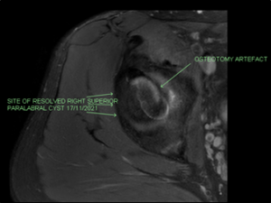
Figure 3b: post-treatment axial MRI demonstrating healing of the labral tear and resolution of the para-labral cysts
Discussion
Corresponding to a clinical study, eighty-seven percent of chronic labral tears without acute trauma demonstrate evidence of femoroacetabular impingement (FAI), hip dysplasia (HD), or osteoarthritis (OA) [5]. Initial management of labral tears consists of activity modification with rest, avoidance of the triggering activities, and physical therapy.
We use pelvic and lower extremity muscle strengthening exercises to stabilize the joint, change the abnormal pelvic tilt, and ease the abnormal pressure on the labrum. If physical therapy fails, arthroscopic surgery is suggested. Given the importance of the labrum for hip joint health, surgery should be focused on restoring the labrum rather than debridement or excision each time possible.
In standard 2D cell culture systems, cells are grown on horizontal dishes with coated shells to support adhering and thriving. These patterns are not typical of the in vivo cell environment as they do not imitate the microenvironment seen in body tissues.
Collagen has been used effectively and safely in non-cultured use in Orthopedics termed Chondro-Gide® as an implant [6] and in rheumatology named ChondroGrid® as a direct injection [7].
In our patient, we used FDA-approved type1 bovine collagen as a medical device using a particular specification of the amino acids in an adequately done trial in vitro. We have combined medical and scientist liaisons. We cross-linked the collagen with hyaluronic acid in 3D stem cell culture. The cells are grown in 3D shapes by cutting the monolayer system of growing cells. 3D cell cultures are complex systems linked together by microfluidics and thus show better cell-ECM and cell-cell interactions.
Mesenchymal stem cells (MSCs) are currently being studied in various research facilities and clinical practices to determine efficacy and safety [8, 9]. In addition, randomized controlled trials showed the positive outcome of MSCs in osteoarthritis [10].
His symptoms have resolved, and an MRI scan showed complete repair of the extensive labral tear and resolution of the para-labral cysts showing a fascinating outcome to 3D stem cell therapy. We have reviewed the relevant literature and found no case in the world like our findings; thus, advancing this technique on a larger scale might be the future mainstream medicine in regenerating the poorly vascular structures like cartilage, labrum, meniscus, tendon, and ligament.
Conclusion
We hypothesize that 3D stem cell culture (hydrogel) has resulted in the complete repair of the extensive labral tear and full resolution of the para-labral cysts. No other non-surgical therapy in the literature has achieved such world excellent results clinically and radiologically within a short period. However, we need to confirm those findings are consistent in broad randomized controlled trials.
References
- Ferguson SJ, Bryant JT, Ganz R, et al. The influence of the acetabular labrum on hip joint cartilage consolidation: a poroelastic finite element model. J Biomech. 2000;33(8):953–960. doi: 10.1016/S0021-9290(00)00042-7.
- Wasielewski RC. The hip. In: The adult hip. Philadelphia: Lippincott-Raven; 1998.
- Bharam S. Labral tears, extra-articular injuries, and hip arthroscopy in the athlete. Clin Sports Med. 2006;25(2):279–292. doi: 10.1016/j.csm.2006.01.003.
- Freehill MT, Safran MR. The labrum of the hip: diagnosis and rationale for surgical correction. Clin Sports Med. 2011 Apr;30(2):293-315.
- Wenger DE, Kendell KR, Miner MR, Trousdale RT. Acetabular labral tears rarely occur in the absence of bony abnormalities. Clin Orthop Relat Res. 2004 Sep.
- Bistolfi A (2017) Regeneration of articular cartilage: Scaffold used in orthopedic surgery. A short handbook of available products for regenerative joint surgery. Clin Sci Res Rep DOI: 10.15761/CSRR.1000101
- Paola De Luca, Alessandra Colombini, et al. Intra-Articular Injection of Hydrolyzed Collagen to Treat Symptoms of Knee Osteoarthritis. A Functional In Vitro Investigation and a Pilot Retrospective Clinical Study. J. Clin. Med. 2019, 8, 975; doi:10.3390/jcm8070975
- Brown MH, Scholes C, Hafsi K, Marenah M, Li J, et al. (2019) Efficacy and safety of culture-expanded, mesenchymal stem/stromal cells for the treatment of knee osteoarthritis: a systematic review. J Orthop Surg Res 14:34.
- Centeno CJ, Al-Sayegh H, Freeman MD, Smith J, Murrell WD, et al. (2018) A multi-center analysis of adverse events among two thousand, three hundred and seventy-two adult patients undergoing adult autologous stem cell therapy for orthopedic conditions. Int Orthop 40: 1755-1765. doi:10.1007/s00264-016-3162-y.
- Han X, Yang B, Zou F, Sun J (2020) Clinical therapeutic efficacy of mesenchymal stem cells derived from adipose or bone marrow for knee osteoarthritis: a meta-analysis of randomized controlled trials. J Comp Eff Res 9: 361-374.
