Publication Information
Frequency: Continuous
Format: PDF and HTML
Versions: Online (Open Access)
Language: English
| Journal Menu |
| Editorial Board |
| Reviewer Board |
| Articles |
| Open Access |
| Special Issue Proposals |
| Guidelines for Authors |
| Guidelines for Editors |
| Guidelines for Reviewers |
| Membership |
| Fee and Guidelines |
 |
An Ectopic Tooth in Nasal Cavity with Nasal Congestion
Bao-Feng Wang, MD, PhD1*; Yi-Jing Chen, MD, PhD1; Ying-Ying Wang, BS1; Yun-Lan Yi, BS2; Ming-Ming Wang, BS1; Xiao-Rui Li, BS3
1Department of Otolaryngology-Head and Neck Surgery, Beijing Tsinghua Changgung Hospital, School of Clinical Medicine, Tsinghua University, Beijing, P.R. China
2Department of Otolaryngology-Head and Neck Surgery, Jinzhou medical College, Jinzhou, P.R. China
3Department of Otolaryngology-Head and Neck Surgery, Minsheng Ear, Nose and Throat Hospital, Wuhan, P.R. China
Received Date: August 02, 2023; Accepted Date: August 14, 2023; Published Date: August 30, 2023;
*Corresponding author: Bao-Feng Wang, Department of Otolaryngology-Head and Neck Surgery, Beijing Tsinghua Changgung Hospital, School of Clinical Medicine, Tsinghua University, Beijing, P.R. China. E-Mail: wangbaofeng2013@aliyun.com
Citation: Wang BF, MD, PhD; Chen YJ, MD, PhD; Wang YY BS, Yi YL, BS; Wang MM BS; Li XR BS (2023) An Ectopic Tooth in Nasal Cavity with Nasal Congestion. Annals of Otorhinolaryngology – Head and Neck: AOHNS-103.
DOI: 10.37722/AOHNS.2023201
Abstract
An ectopic tooth within the nasal cavity is an exceedingly rare and unusual occurrence within the realm of the otolaryngology department, which focuses on ear, nose, and throat medicine. Here, we present a remarkable case of a nasal cavity ectopic tooth resulting in three months of left nasal obstruction, detected during an examination conducted with an electronic nasopharyngoscope. The tooth was found to be protruding from the floor, situated in the inferior nasal meatus, accompanied by the presence of granulation tissue. The diagnosis was unequivocally confirmed as an ectopic tooth lodged in the nasal cavity. Detacher extraction was performed under general anesthesia. The objective of this study was to gain insights into the etiology, diagnostics, and therapeutic approaches associated with ectopic teeth.
Keywords: ectopic tooth, nasal cavity, tooth extraction
Introduction
An intranasal retained tooth is an uncommon occurrence, with an estimated incidence ranging from 0.1% to 1% in the general population [1]. Ectopic teeth is a disturbance in which the tooth does not follow its usual course, and can be found in various regions within the nose, with the majority located in the common meatus [2].However, the rarest situation involves their placement in the inferior nasal meatus. The presence of common nasal symptoms such as congestion, epistaxis, and discharge often raises suspicion for chronic rhinosinusitis; [3, 4] nonetheless, it is crucial to consider a differential diagnosis.
Given the rarity of intranasal retained teeth, there is a paucity of literature on this subject, making it a relatively unexplored area of research. Due to their potential mobility, a prompt and accurate diagnosis becomes of utmost importance in managing such cases.
Case presentation
A 29-year-old male patient presented to the otorhinolaryngology department with a three-months history of left-sided nasal congestion and persistent purulent discharge. Despite using topical nasal steroids for a month, there was no noticeable improvement in his condition.
The patient denied any previous nasal or craniofacial bone injuries. An endoscopic examination revealed a deviated septum towards the left side and an accompanying ipsilateral bone spur (Figure 1).
To address the condition, we performed a detacher removal procedure to remove the bone spur (Figure 2). Additionally, the patient was instructed to use nasal saline spray, which was also prescribed to aid in his recovery.
Discussion
As previously mentioned, an atopic tooth in the nasal cavity is a rare occurrence, with a higher incidence in males than females.6 The most frequent occurrence of an ectopic tooth typically manifests in the upper incisor region, which is referred to as the mesiodens [5].This condition can give rise to a variety of symptoms, including but not limited to pain in the nasal area, nasal discharge, nasal obstruction, rhinorrhea, headache, and epistaxis.6-7 Although the exact cause remains unclear, it has been theorized to be linked to developmental abnormalities, facial traumas, or overcrowding of the teeth leading to abnormal eruption, or genetic factors [6, 7, 8]. Conditions like gardeners and cleidocranial dystosis have also been associated with the development of an ectopic tooth [9]. In our case there was no obvious explanation of the etiology.
Upon examination, patients with this condition typically present with a unilateral nasal mass covered by mucosa, nasal secretions, and granulation tissues. The differential diagnosis may consider foreign body, rhinolith, bony sequestration, and even neoplasm, making it crucial to conduct careful examinations [10]. And endoscopic examinations and radiological investigations, such as computerized tomography (CT) scan for paranasal sinuses, play a significant role in aiding the diagnosis [9]. These investigations may reveal a radiopaque mass, and in some cases, they can even confirm the diagnosis, as was the case with our patient.
Urgent treatment is crucial as the presence of the ectopic tooth can cause significant discomfort to the patient and lead to various complications as upper mentioned. The recommended treatment is the extraction of the tooth, typically performed through an endoscopic view trans-nasal approach, because it ensures clear visualisation and is minimally invasive [3]. Care must be taken to ensure complete and cautious removal of the tooth to avoid further mucosal injury. This procedure can be carried out in the clinic under local anesthesia if the patient is cooperative or in the operating room under general anesthesia. Our case report demonstrates that the surgical approach employed is both straightforward and safe, effectively resolving all symptoms.
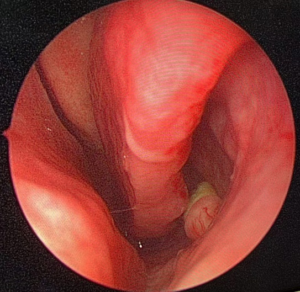
Figure 1: a tooth covering with granulation in the root in the left inferior nasal meatus.
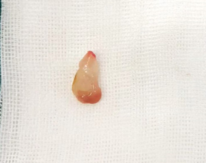
Figure 2: the removal tooth from nasal cavity. The dimensions of it were 1.0 cm * 0.4 cm * 0.2 cm. cm, centimeter.
Reference
- Bergamaschi IP, Olsson B, Sebastiani AM, Trento GDS, Rebellato NLB, Klüppel LE, et al. Intranasal Ectopic Tooth in Adult and Pediatric Patients: A Report of Two Cases. Case Rep Surg. 2019;2019:8351825.
- Almomen A, Alkhudair B, Alkhatib A, Alazzah G, Ali Z, Yaeesh IA, et al. Ectopic maxillary tooth as a cause of recurrent maxillary sinusitis: a case report and review of the literature. J Surg Case Rep. 2020; 2020(9): rjaa334.
- Gupta S, Cipolla M. Endoscopic Surgical Excision of Ectopic Tooth in Left Nasal Cavity. Cureus. 2021; 13(8): e17465.
- Gormley M, Chahal R, Gallacher N and Bell C. A conventional surgical approach for removal of an ectopic tooth in the nasal cavity. BMJ Case Rep. 2019; 12(8): e231279.
- Kratz B, Chhabra N. Ectopic Tooth of the Nasal Cavity. Ear Nose Throat J. 2020;99(2):NP13-NP14.
- Ahmed AK, Alansari H, Almannai A, Ali HY. Should we floss our nose? A case report of a tooth in the nasal cavity. Int J Surg Case Rep. 2021; 80: 105664.
- Alfayez N, ALHumaid SA and Alfayez A. Ectopic nasal tooth: A case report. Int J Surg Case Rep. 2021; 88:106459.
- Peck S, Peck L, Kataja M. The palatally displaced canine as a dental anomaly of genetic origin. Angle Orthod. 1994;64:249–256.
- Subasioglu A, Savas S, Kucukyilmaz E, Kesim S, Yagci A, Dundar M. Genetic background of supernumerary teeth. Eur J Dent. 2015; 9(1):153-8.
- Raubaite R, Rakauskaite A, Sukyte-Raube D, Zaleckas L, Rauba D. An Intranasal Ectopic Tooth in an Adult. Cureus. 2022;14(4):e24410.
